Problem with lung motion in radiation therapy and current 4D methods
Technology
5DCT brings accurate tumor delineation and treatment planning
Patients with inconsistent breathing patterns complicate 4DCT acquisition. Even with coaching, some patients may fail to produce regular breathing traces, increasing the likelihood of motion-related inaccuracies in imaging and treatment delivery.
Our product, Repirati 5D®
Our product, Respirati 5D®, acquires and analyzes CT images using an innovative model-based approach that provides the user with images at user-selected breathing phases to display an accurate reflection of tumors that move during breathing. Our product replaces the existing commercial system which suffers from image errors and sensitivity to irregular breathing patients. The reconstructed images provided by the product will increase the accuracy of radiation therapy treatment planning optimization and delivery of radiation therapy to cancer patients.
Our 5D solution – Meet Respirati 5DCT
We start with a series of conventional helical CT scans while monitoring breathing patterns, that are generally irregular. This provides us with multiple breath-hold-like images sampled over two minutes (to sample multiple breathing cycles) that are compared with each other (using deformable image registration) to characterize the tumor motion and correlating it with the breathing amplitude and rate. We combine this tumor motion information (x, y, and z), with the breathing information (amplitude and rate) to generate artifact-free images for treatment planning. Respirati 5DCT has been shown to outperform all commercially available image reconstructions methods to prepares the radiation oncology team with the most accurate tumor and motion margin (ITV) delineation for treatment planning.
The 5DCT name stems from the motion model’s description of tissue displacement as a function of the five variables: x, y, and z position, breathing amplitude, and breathing rate.

4DCT
Limitations of 4DCT methods in radiation therapy
Despite being the most widely used technology for reconstructing lung tumor motion, 4DCT methods do not accurately measure tumor volume and breathing motion. The main reason is that most patients breathe irregularly, leading to erroneous sampling and subsequent image artifacts.
As a result, 4DCT images often misrepresent tumor volumes.
5DCT®
Respirati 5DCT® combines a novel CT scanning technique and a motion model to provide artifact-free images and accurate tumor volume and motion for patients who breathe irregularly.
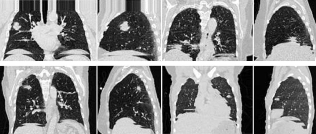
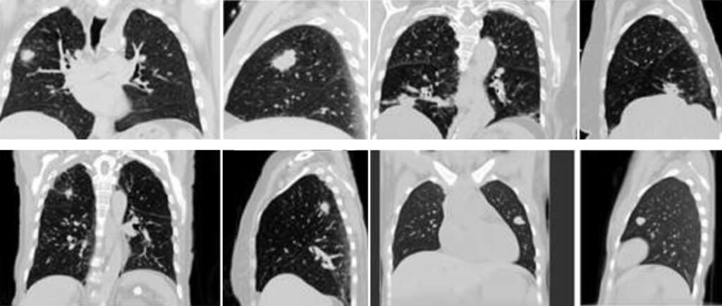
Simulated cone-beam CT scan of a free-breathing 5DCT patient. The motion model, along with their breathing pattern, is used to generate this simulated 5DCT scan. Similar work can be done simulating treatment delivery.
FAQs
FAQs about the limitations of 4DCT
Irregular breathing can cause artifacts in the images, such as blurring, duplication, or missing anatomy. This makes it harder to accurately define tumor motion and may affect treatment planning. Coaching the patient beforehand or using motion management systems can help minimize this.
No, 4DCT is designed to track respiratory motion. It does not capture motion caused by other factors, like heartbeat, digestion, or posture changes, which may also affect tumor location in certain body sites.
The temporal resolution of 4DCT depends on the CT scanner’s speed and the patient’s breathing rate. Very fast or irregular breathing may lead to incomplete motion capture or distortion of anatomy at specific phases of the cycle.
Binning is the process of dividing scan data into different phases of the breathing cycle. If the sorting algorithm or patient’s breathing is inconsistent, it can result in misassigned images, leading to inaccurate reconstructions of tumor and organ positions.
Not always. Patients who cannot follow breathing instructions, such as those with cognitive impairment, anxiety, or respiratory illness, may not tolerate the scan well, resulting in poor-quality images or the need for a repeat scan.
Because 4DCT reconstructs multiple image sets from a single breathing cycle, individual phases often have lower signal-to-noise ratio and reduced spatial resolution, making small lesions or soft tissue boundaries harder to distinguish.
Yes, 4DCT typically delivers a higher radiation dose than a standard 3D CT because it captures images across the entire breathing cycle. However, the clinical benefits usually outweigh the risks, especially for accurate treatment planning.
Not exactly. 4DCT captures motion during a single scan session, which may not represent day-to-day variations in breathing or tumor position. To address this, some clinics use daily imaging, motion management systems, or adaptive planning techniques.
Yes. Breathing coaching, audio-visual feedback systems, and advanced motion management tools can improve scan quality. Some centers also use MRI or PET/CT as complementary imaging for better soft tissue contrast and motion assessment.
FAQs about 5DCT versus 4DCT
5DCT stands for 5-dimensional computed tomography. Like 4DCT, it captures breathing-related motion, but it adds a statistical modeling layer to generate a more accurate and continuous representation of anatomy over time.
- 4DCT shows motion in a few discrete breathing phases.
- 5DCT reconstructs motion as a continuous function, accounting for both amplitude and phase variations in respiration—hence the “5th dimension.”
5DCT uses many more projections collected over multiple breathing cycles and applies motion modeling techniques. This results in:
- Sharper images with fewer motion artifacts
- Higher spatial and temporal resolution
- More consistent anatomy across the full breathing range
Yes. Unlike 4DCT, which suffers from phase-sorting errors due to inconsistent breathing, 5DCT builds a statistical model of motion across the entire scan, making it more robust to irregular breathing patterns and reducing artifacts.
While both are typically single-session scans, 5DCT provides a more flexible and realistic model of breathing motion by not relying on rigid phase binning. This makes the motion envelope it creates more reflective of what might occur across treatment days.
Yes. 5DCT provides a continuous motion model, allowing better estimation of the Internal Target Volume (ITV) and reducing the risk of over- or under-estimating motion. This leads to more precise Planning Target Volume (PTV) margins.
With higher image fidelity and more realistic motion modeling, 5DCT allows for:
- More accurate dose calculations
- Better adaptation to motion
- Improved organ-at-risk sparing This enhances the overall quality of the treatment plan.
The scanning process is comparable or slightly longer, but the reconstruction and processing time is more involved due to advanced motion modeling. However, automated software tools are improving to streamline clinical workflows.
The dose is similar or slightly higher than 4DCT depending on scanner settings. However, the improved motion representation can lead to better treatment accuracy, potentially allowing smaller margins and less total dose to healthy tissues.
5DCT is especially useful in:
- Thoracic and upper abdominal tumors
- Patients with irregular or shallow breathing
- Cases where precise motion characterization is critical for planning or adaptive therapy
Not yet. 5DCT is still emerging and only available at UCLA Health clinics. It will soon be available for broader distribution.
Comparisons of 4DCT vs 5DCT for Radiation Therapy
| Feature | 4DCT | 5DCT |
|---|---|---|
| What it is | CT scan over the respiratory cycle, divided into discrete breathing phases | CT scan with advanced motion modeling over time and amplitude—continuous representation |
| Breathing Motion Capture | Discrete bins (typically 8–10 phases) | Continuous, model-based motion envelope |
| Image Quality | Susceptible to motion artifacts and sorting errors | Higher fidelity, less noise, and reduced artifacts |
| Handling Irregular Breathing | Prone to errors and artifacts | Robust modeling handles inconsistencies better |
| Temporal Resolution | Limited by bin count and scanner speed | Statistically modeled—more accurate across time |
| Motion Modeling | Phase-based snapshots only | Captures both phase and amplitude variations (5th dimension) |
| Tumor Margin Accuracy | Risk of over- or underestimating ITV | More accurate ITV/PTV definition |
| Dose Calculation | Based on selected breathing phases | Reflects full motion range—improved accuracy |
| Radiation Exposure | Moderate to high | Comparable, possibly slightly higher |
| Ease of Use | Widely available and supported | Emerging technology—requires advanced software |
| Clinical Suitability | Most patients with predictable breathing | Best for patients with variable or shallow breathing |
| Availability | Standard in most radiation oncology clinics | Limited to research or advanced centers (for now) |